Skip to content
Identify various parts of the nervous system, including the brain, spinal cord, and nerves.Discuss the necessity of the spinal cord, brain, and nerves for the body.Understand how all parts of the body are connected to the brain through nerves.List activities that are under the control of the nervous system.
Nervous System: Exercises neural control through electrochemical impulses.Endocrine System: Exercises chemical control through chemical messengers called hormones.
Coordination: Normal control over the functioning of body tissues and organs at the correct speed and in the correct sequence simultaneously within physiological limits.Example of Coordination (Eating Food): Involves looking at and identifying food items, putting the food on a plate, using a spoon or fork, chewing, swallowing, digestion, and absorption—all in a coordinated sequence using different organs.Stimulus: Information received by the nervous system about the conditions in the environment inside or outside the body.Response: A reaction to the stimulus; the behavior shown by a living organism as a result of an external or internal stimulus.

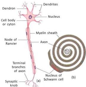
Cell Body or Cyton: The main part of a neuron containing the nucleus. It receives impulses from the preceding neuron through dendrites and passes them to the next neuron through the axon.Dendrons and Dendrites: Several finger-like projections branching out from the cyton are called dendrons. The fine branches of dendrons are called dendrites, which end in knob-like structures called terminal knobs. Dendrites connect a neuron with neighboring neurons, receiving impulses and conducting them to the cyton (Afferent process).Axon: The longest single extension from the cyton. Its end is divided into terminal branches that end in synaptic knobs. The axon and its terminal branches transmit impulses away from the cyton to the dendrites of other neurons or to an effector organ (Efferent process).
In most neurons, the axons are surrounded by a spirally-coiled sheath of a fatty substance called the myelin sheath (or modulated sheath).Function: The myelin sheath insulates the axon and speeds up the conduction of nerve impulses. This explains why the speed of nerve impulses is higher in myelinated nerve fibers compared to non-myelinated ones.Nodes of Ranvier: Regular constrictions along the axon where the myelin sheath is interrupted.Schwann Cell: The myelin sheath contains the nucleus of a Schwann cell.
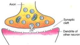
The point of contact between the terminal branches of the axon of one neuron and the dendrites of an adjacent neuron is called a synapse.At the junction, there is a fluid-filled gap that separates the neurilemma of the two neighboring neurons.The nerve impulse passes through the synapse as a chemical message.This propagation is carried out by chemical messengers called neurotransmitters. The primary neurotransmitter at the nerve synapse is acetylcholine.
Nerve Fibers: These are individual axons, some of which can be more than a meter long.Nerve: A bundle of nerve fibers wrapped together in a protecting sheath of connective tissue. This sheath keeps individual nerve fibers isolated inside the nerve.Analogy: A nerve can be compared to an electric cable, where the entire cable is the nerve and the internal insulated wires are the individual nerve fibers (axons) protected by their sheaths.

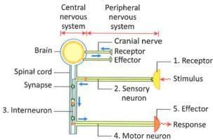
Receiving the Stimulus: Sensory cells or receptors receive stimuli caused by changes in either the external or internal environment.Processing of Stimulus (Transduction): Receptors convert physical or chemical stimuli into electrical nerve impulses.Transmission of Nerve Impulse: The receptors transmit these electrical sensory impulses to the sensory neurons present in the brain or spinal cord.Interpretation of Sensory Impulses: Interneurons in the brain or spinal cord interpret the incoming sensory impulses and convert them into outgoing motor impulses.Transferring the Response (Action): Motor nerve fibers (axons of motor neurons) transfer the response from the brain or spinal cord to the effectors (muscles or glands). The effectors act according to the orders, translating the stimulus into a physical body response.
Central Nervous System (CNS): Consisting of the brain and spinal cord. It plays an integration role, processing information and instructing effectors on how to respond.Peripheral Nervous System (PNS): Consisting of cranial and spinal nerves that connect the CNS to all parts of the body.
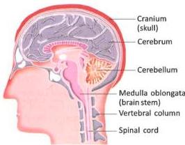
Protection: It is protected by three protective membranes called meninges and is enclosed inside a bony brain case called the cranium (or skull).Tissue Composition:Gray Matter: Forms the outer layer of the brain and is mainly composed of cell bodies of neurons.White Matter: Forms the inner layer of the brain and contains myelinated axons that group to form nerves.CerebrumCerebellumMedulla Oblongata
Structure:It is the largest and most highly developed part of the brain.It contains an estimated nine billion neurons, which explains its capacity to process immense volumes of cognitive tasks.It is divided into right and left halves called cerebral hemispheres by a deep groove.Each hemisphere is hollow, and the outer layer of gray matter is highly folded into ridges called gyri and grooves called sulci to accommodate a massive number of neurons.Functions:It is the seat of intelligence, consciousness, and willpower.It controls cognitive functions like reasoning, instinct, thinking, learning, memorizing, and complex emotions (love, hatred, appreciation).It is responsible for sensory perceptions of sight, hearing, taste, smell, pain, pressure, temperature, and touch.
Structure: Located at the back of the cerebrum and is partly overlapped by it.Functions:Coordinates voluntary movements of the body.Controls the activities of voluntary muscles.Maintains body posture and physical balance during active states like walking, swimming, jumping, and running.
Structure: It is the posterior-most part of the brain that connects to the spinal cord.Functions:Coordinates all involuntary activities of internal (visceral) organs.Regulates critical vital processes such as breathing movements, heartbeat, swallowing, and peristaltic movements of the alimentary canal.Critical Note: Any injury to the medulla oblongata can be immediately fatal, resulting in the death of a person due to the stoppage of vital involuntary functions.
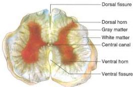
Structure:It extends downward from the medulla of the brain up to the lumbar region of the lower back.It runs through the protective neural canal of the vertebral column (spine), which protects it from physical injury.Tissue Arrangement: The arrangement of gray and white matter in the spinal cord is the reverse of the brain:Gray Matter: Centrally located and forms an H-shaped region.White Matter: Forms the outer peripheral layer.Functions:Controls all types of reflex actions (automatic, instantaneous responses to stimuli independent of voluntary control).Mediates involuntary visceral activities such as heartbeat, secretion of glands, and breathing movements.Serves as a conduction pathway, carrying sensory impulses from the body to the brain and returning motor impulses from the brain to effectors.
Cranial Nerves: 12 pairs of nerves that arise directly from the brain and supply different parts of the head.Spinal Nerves: 31 pairs of nerves that arise from the spinal cord and supply different parts of the body excluding the head.Composition: Every spinal nerve is a mixed nerve, containing both sensory and motor nerve fibers.
Structure: It consists of paired chains of ganglia (clusters of nerve cell bodies) lying on either side of the vertebral column, which are connected directly to the spinal cord.Divisions: The ANS is divided into two antagonistic systems that produce opposite reactions in the same target organs:Sympathetic Nervous SystemParasympathetic Nervous System
Share
Explore
 Self Study
Prepared by: learnloophq@gmail.com
Chapter: 07. Nervous System
7 Nervous System
Learning Outcomes
After studying these notes, you will be able to:
Section 1: Control and Coordination
Multicellular organisms have evolved two coordinated systems to ensure efficient control and coordination of various activities occurring simultaneously in different body systems:
Key Concepts in Coordination
Pathway of Nervous Coordination
The pathway of an impulse from a stimulus to a response follows this sequence:

Section 2: Neurons and Nerve Structure
The nervous system acts as the control center of the body. It receives stimuli from inside and outside the body through receptors and five sense organs: eyes, ears, nose, tongue, and skin. It is made of a network of nerve cells (neurons) and nerve fibers, providing the fastest means of communication between different body parts.
Structure of a Neuron (Nerve Cell)
Neurons are the structural and functional units of the nervous system. The nervous system is made of millions of neurons. A neuron consists of three main parts:
Myelin Sheath
Synapse
Nerve Fibers and Nerves
Section 3: Types of Nerves and Mechanism of Coordination
Types of Nerves
Nerves are classified into three types based on the nature of the impulses they conduct:
Type of Nerve
Composition
Function
Examples
Sensory Nerves
Only sensory nerve fibers
Bring impulses from sense organs to the brain or spinal cord
Optic nerve of the eye
Motor Nerves
Only motor nerve fibers
Carry impulses from the brain or spinal cord to effector organs (muscles/glands)
Nerves supplying eye muscles and muscles of the tongue
Mixed Nerves
Both sensory and motor nerve fibers
Transmit both incoming sensory and outgoing motor impulses
Nerves going to the lower jaw, face, neck, and salivary glands
Mechanism of Nervous Coordination
Nervous coordination operates in five distinct sequential steps:

Section 4: Human Nervous System and Central Nervous System (CNS)
The human nervous system is split into two primary divisions:
(Note: The Autonomic Nervous System is also part of the broader nervous network, managing involuntary visceral activities.)

1. The Brain
The brain is the primary controlling part of the nervous system.
The brain consists of three main parts:
A. Cerebrum
B. Cerebellum (“Little Brain”)
C. Medulla Oblongata (“Brain Stem”)
2. The Spinal Cord

Section 5: Peripheral and Autonomic Nervous Systems
1. Peripheral Nervous System (PNS)
The PNS consists of all the nerves that branch out from the brain and spinal cord, serving to connect the CNS to every part of the human body.
2. Autonomic Nervous System (ANS)
The ANS controls the involuntary actions of visceral organs and the activities of smooth muscles in the alimentary canal and blood vessels.
Differences Between Sympathetic and Parasympathetic Nervous Systems
Feature
Sympathetic Nervous System
Parasympathetic Nervous System
Origin of Nerves
Arise from the thoracic and lumbar regions of the spinal cord.
Arise from the brain, cervical, and sacral regions of the spinal cord.
Ganglia Location
Form a chain running parallel and close to the spinal cord.
Lie close to or inside the target organ.
General Effect
Has a stimulatory effect on body organs.
Has an inhibitory or calming effect on body organs.
Functional Role
Prepares the body for violent action against adverse situations (“fight-or-flight”).
Brings body activities back to normal state (“rest-and-digest”).

Want to print your doc?
This is not the way.
This is not the way.

Try clicking the ··· in the right corner or using a keyboard shortcut (
CtrlP
) instead.