Skip to content
Analogy to a Machine: A living organism functions similarly to a complex machine. Just as a machine has different parts designed to execute specific jobs, living organisms possess specialized body parts that work in an organized, coordinated fashion.Definition of Organisation: Organisation is the precise manner in which the smaller units of any structure or system are arranged into larger units in a hierarchical fashion.Purpose of Organisation: In multicellular organisms, organisation is beneficial as it enables the division of labour. This division helps cells, tissues, and organs carry out distinct life functions more efficiently and in a highly coordinated manner.
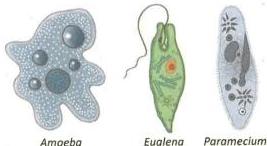
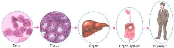
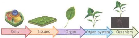
Characteristics: The cell is the smallest, basic living unit that can exist independently and perform all life activities.Unicellular Organisms: In organisms consisting of a single cell—such as Amoeba, Euglena, Paramecium, and bacteria—all life processes (nutrition, respiration, excretion, and reproduction) are carried out within that single cell.Simple Multicellular Organisms: Simple organisms like algae and sponges represent a basic cellular level of organisation. While their cells are grouped together, they function independently and lack a true division of labour.Beginning of Division of Labour: In complex multicellular organisms, cells become structurally specialized to perform specific tasks.Nerve cells: Specialised to conduct messages.Muscle cells: Specialised for contraction and relaxation.Red Blood Cells (RBCs): Specialised for transporting oxygen.Gland cells: Specialised for secretion.Definition of Tissue: A tissue is a group of structurally similar cells that perform a similar function.Characteristics: Different tissues that work closely together to perform a specific, specialized function form an organ.Examples in Animals: Brain, kidney, stomach, intestine, lungs, and heart.Examples in Plants: Root, stem, leaf, and flower.Characteristics: A group of different organs working together in a coordinated manner to perform a major life function forms an organ system.Examples in Animals:Digestive System: Formed by the mouth, foodpipe, stomach, intestine, and rectum.Circulatory System: Formed by the heart and blood vessels.Examples in Plants: Plants have two primary organ systems:Shoot System: Consists of the stem, branches, leaves, flowers, and fruits.Root System: Consists of the main root, its branches, and the root hairs.Characteristics: At this level, all the organ systems function together in a highly coordinated, integrated manner, allowing the individual multicellular organism to survive and interact with its environment.
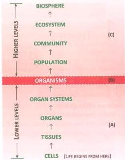
Population Level: Includes all individuals of a particular species living and interacting within a specific geographical area.Community Level (Biotic Community): Consists of populations of different species living and interacting together in a specific area.Ecosystem Level: Formed by the interaction between the members of a biotic community and the abiotic components of their environment.Interdependence among organisms for food establishes interconnected food chains and food webs.Energy Transfer: Energy is transferred sequentially from organisms of one trophic level to those of another level within the food chain.Biosphere Level: The entire inhabited part of the Earth where living organisms exist.
Cell Shape and Size: Cells are typically small and oval or polygonal.Cell Wall: Extremely thin cell walls.Intercellular Spaces: Cells are tightly packed with no intercellular spaces between them.Nucleus: Large and centrally located.Vacuoles: Either completely absent or exceptionally small.Apical Meristem: Located at the growing points or tips of roots, shoots, and branches. It is responsible for the increase in the length of the plant.Lateral Meristem: Located on the lateral sides of stems and roots. It is responsible for the increase in the thickness or girth of the stem and root.Facilitate primary growth (length/height) and secondary growth (girth/thickness) in plants.Give rise to all new plant organs such as buds, flowers, new leaves, and branches through active cell division.
Derived directly from the division of meristematic cells.Cells do not divide because they have undergone differentiation.Cells may be alive or dead.They possess varied shapes that directly correlate with the functions they perform.Cells exhibit thick cell walls and contain a large central vacuole.The nucleus is displaced to one side of the cell.
Epidermis: Forms a continuous single layer of cells covering the entire outer surface of the plant.Cuticle: The epidermis of leaves secretes a waxy, waterproof external layer called the cuticle to prevent excessive water loss.Cork: Consists of dead, tightly packed cells. It is found exclusively in old, woody dicot stems, replacing the epidermis to provide robust protection.Parenchyma:Structure: Found in the soft parts of the plant. The cells are oval, round, or polygonal, with thin cell walls and large vacuoles. They are loosely arranged, leaving distinct intercellular spaces between them.Chlorenchyma: Specialized parenchyma cells containing chlorophyll that perform photosynthesis.Functions: Store food materials, form ground tissue, assist in photosynthesis (in leaves), and facilitate the conduction of water.Collenchyma:Structure: Composed of elongated, living cells. Their cell walls are unevenly thickened, particularly at the corners. Consequently, there are no intercellular spaces. Found beneath the epidermis in leaves, stems, and petioles of herbaceous dicot plants.Functions: Provides mechanical support, elasticity, and flexibility to leaves and stems, preventing them from breaking easily under strain.Sclerenchyma:Structure: Consists of long, narrow, dead, and fiber-like cells. They possess extremely thick, lignified cell walls, leaving a very small central cavity (lumen). They contain no cytoplasm or nucleus at maturity. Located around vascular bundles in stems, in the veins of leaves, and in petioles.Functions: Provides exceptional mechanical strength, rigidity, and stiffness to the plant body.
Function: Responsible for the upward transport of water and dissolved minerals from the roots to the leaves and other aerial parts of the plant.Composition: Composed of four distinct types of cells:Xylem Tracheids: Elongated, dead, tube-like cells with tapering ends.Xylem Vessels: Long, tubular structures formed by dead cells joined end-to-end like drain pipes, with their end partitions dissolved to form continuous channels.Xylem Fibres: Elongated, thick-walled dead cells providing mechanical support.Xylem Parenchyma: The only living cells in xylem tissue; involved in lateral conduction and storage.Note: Because older, non-functional xylem forms the tough wood of trees used in furniture, xylem is commonly referred to as wood.Function: Responsible for the translocation (downward and lateral transport) of prepared organic food from the leaves to all other growing and storage regions of the plant.Composition: Composed of four distinct types of cells:Sieve Tubes: Long, tubular conducting channels joined end-to-end. Their end-plates are perforated with fine pores and are known as sieve plates to allow easy passage of food.Companion Cells: Living cells closely associated with sieve tubes, assisting in their function.Phloem Parenchyma: Living cells that store food materials.Phloem Fibres: The only dead cells in phloem tissue; provide mechanical support.
Share
Explore
 Self Study
Prepared by: learnloophq@gmail.com
Chapter: 01. Tissues
Chapter 1: Tissues
SECTION 1: INTRODUCTION TO ORGANISATION & LEVELS OF ORGANISATION
Concept of Organisation
Levels of Organisation Lower to the Organism
[CELLS] ➔ [TISSUES] ➔ [ORGANS] ➔ [ORGAN SYSTEMS] ➔ [ORGANISMS]
1. Cellular Level
2. Tissue Level
Know Your Scientist: Marie Francois Xavier Bichat Marie Francois Xavier Bichat was a French anatomist and pathologist. He introduced the term tissue in the 180s (as noted in the historical record). Bichat proposed that tissues are the central elements in human anatomy and that organs are collections of different tissues. For these pioneering contributions, he is recognized as the Father of Modern Histology.
3. Organ Level
4. Organ-System Level
Levels of biological organisation in animals
Levels of biological organisation in animals
Levels of biological organisation in plants
Levels of biological organisation in plants
5. Organism Level
Levels of Organisation Higher to the Organism
No living organism exists in absolute isolation. It interacts continuously with both the living (biotic) and nonliving (abiotic) components of its environment. This gives rise to higher levels of organization:
Section 1: Visual Recap

SECTION 2: PLANT TISSUES
Plant tissues are broadly classified into two major categories based on their capability to divide: Meristematic Tissues and Permanent Tissues.
PLANT TISSUES
│
┌──────────────────────┴──────────────────────┐
Meristematic Tissues Permanent Tissues
│ │
┌─────┴─────┐ ┌─────────────┴─────────────┐
Apical Lateral Simple Complex
│ │
┌──────────┴──────────┐ ┌───┴───┐
Protective Supporting Xylem Phloem
│ │
┌───┴───┐ ┌──────┼──────┐
Epidermis Cork Parenchyma │ Sclerenchyma
Collenchyma
Meristematic Tissues (Meristems)
Meristematic tissues consist of actively dividing, young, undifferentiated cells. They continuously add new cells to the plant body throughout the plant’s life and are directly responsible for growth.
1. Characteristics of Meristems
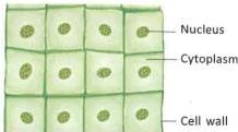
Cells of meristematic tissue
Cells of meristematic tissue
2. Location and Types of Meristems
3. Functions of Meristematic Tissues
Permanent Tissues
Permanent tissues are formed from mature, differentiated cells derived from meristematic tissue. Once cells mature, they lose their ability to divide and specialize to perform distinct roles.
1. Characteristics of Permanent Tissues
2. Key Differences between Meristematic and Permanent Tissues
Meristematic Tissues
Permanent Tissues
1. Cells are young and undifferentiated.
1. Cells are mature and fully differentiated.
2. Cells retain the power to divide continuously.
2. Cells lose the ability to divide.
3. Cells are small, lacking intercellular spaces and vacuoles.
3. Cells can be living or nonliving; living cells have intercellular spaces and vacuoles.
4. Cell walls are thin.
4. Cell walls are thick.
Types of Permanent Tissues
Permanent tissues are divided into two main categories: Simple Permanent Tissues and Complex Permanent Tissues.
A. Simple Permanent Tissues
Formed of only one type of cell. They are classified into Protective and Supporting tissues based on their functions.
1. Protective Tissues
Formed of thick-walled cells designed to protect the internal parts of plants.
2. Supporting Tissues
These tissues provide mechanical support, flexibility, and structural strength to the plants.
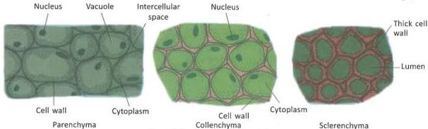
Types of supporting tissues: Parenchyma, Collenchyma, and Sclerenchyma
Types of supporting tissues: Parenchyma, Collenchyma, and Sclerenchyma
3. Key Differences between Parenchyma, Collenchyma, and Sclerenchyma
Feature
Parenchyma
Collenchyma
Sclerenchyma
Intercellular Spaces
Cells are loosely packed with abundant intercellular spaces.
Very little intercellular space between the cells.
No intercellular spaces between the cells.
Cell Walls
Thin cell walls.
Cell wall is irregularly thickened at the corners.
Cell wall is uniformly thick and lignified.
Distribution
Widely distributed in soft parts of the plant body.
Located below the epidermis of herbaceous stems and petioles.
Found around vascular bundles, leaf veins, and petioles.
Vital State
Cells are living.
Cells are living.
Cells are dead.
B. Complex Permanent Tissues (Conducting/Vascular Tissues)
Complex permanent tissues are made of more than one type of cell working together as a single unit to perform a common function. They form the vascular or conducting channels of the plant.
1. Xylem (also known as Wood)
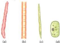
Cell types found in xylem tissue: (a) xylem tracheid, (b) xylem vessel, © xylem fibre, and (d) xylem parenchyma
Cell types found in xylem tissue: (a) xylem tracheid, (b) xylem vessel, © xylem fibre, and (d) xylem parenchyma
2. Phloem
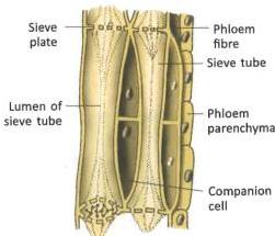
Cell types found in phloem tissue showing sieve tubes, sieve plates, companion cells, phloem parenchyma, and phloem fibres
Cell types found in phloem tissue showing sieve tubes, sieve plates, companion cells, phloem parenchyma, and phloem fibres
3. Key Differences between Simple and Complex Permanent Tissues
Simple Permanent Tissues
Complex Permanent Tissues
1. Formed of only one type of cell.
1. Formed of more than one type of cell.
2. All cells in the tissue perform the same function.
2. Different cell types carry out distinct sub-functions to achieve a single major goal.
3. Three types: Parenchyma, collenchyma, and sclerenchyma.
3. Two types: Xylem and phloem.
4. Primarily provide protection and structural/mechanical support.
4. Primarily transport water, minerals, and synthesized food sugars.
Section 2: Visual Recap

SECTION 3: ANIMAL TISSUES
Want to print your doc?
This is not the way.
This is not the way.

Try clicking the ··· in the right corner or using a keyboard shortcut (
CtrlP
) instead.