Chapter: 06. Human Circulatory System
6 Human Circulatory System
Learning Outcomes
Children will be able to:
Explain the main parts of the circulatory system. List the components of blood and types of blood vessels. Take their own/others’ pulse. Demonstrate the significance of exercise and good food habits in keeping the heart healthy. Introduction to Human Circulatory System
The circulatory system acts as the transport system in our body. Its primary functions include:
Providing body cells with food and oxygen. Removing waste products from body cells and transporting them to the kidneys. Connecting all the cells, tissues, and organs of the body through a system of vessels. Key Concepts Overview
The human circulatory system involves several key components and processes:
Human Circulatory System Parts: Heart - The pumping organ Blood Groups and Blood Transfusion: Antigen and antibody interaction Blood donation and blood bank Keeping the heart healthy There are three main parts of the circulatory system: blood, blood vessels, and heart.
Blood
Blood is a red-coloured, viscous fluid that flows through blood vessels, reaching every cell of the body.
An average human body contains 4.7 to 5.5 litres of blood. Components of Blood
Blood consists of two main components: Plasma and Blood corpuscles (or blood cells).
The liquid part of blood. Makes up more than half (55%) of blood volume. Contains dissolved nutrients, oxygen, enzymes, hormones, waste products, etc. Blood Corpuscles: Blood contains three kinds of blood corpuscles: Red blood corpuscles (RBCs), White blood corpuscles (WBCs), and Blood platelets. Red blood corpuscles (RBCs) or Erythrocytes: Most numerous cells in blood. Approximately 5 million per mL in men, 4.5 million per mL in women. Lack a nucleus (enucleated). Cytoplasm contains haemoglobin, an oxygen-carrying pigment that gives blood its red colour. Haemoglobin combines with oxygen to form oxyhaemoglobin, which transports oxygen to all body cells. New RBCs are formed in the bone marrow. White blood corpuscles (WBCs) or Leucocytes: Have oval or lobed nuclei. Approximately 5000 to 6000 per mL of blood. Defend the body against diseases. Fight against germs and provide immunity against infection. Often called “soldiers of the body.” WBC count increases during infection. Blood platelets or Thrombocytes: Somewhat rounded and very minute fragments of cells. Number 2-5 lakhs per mL of blood. Help stop bleeding at injury sites by clotting the blood.
Fig. 6.1 Blood corpuscles in human blood Functions of Blood
Blood performs several vital functions in the body:
Transports digested food and oxygen to every cell of the body. Removes carbon dioxide from cells and transports it to the lungs. Removes wastes from cells and takes them to the kidneys. Helps in regulating body temperature. Protects the body against infections. Transports hormones to all body parts. Helps in clotting of blood and healing of wounds. Maintains the balance of salt (sodium) and water in body fluid and tissues.
Blood Vessels
Blood flows through a system of tubes called blood vessels. There are three kinds of blood vessels: Arteries, Veins, and Capillaries.
Arteries
Distributing vessels: Carry blood away from the heart to other parts of the body. Have thick and muscular walls. Generally carry pure or oxygenated blood (containing oxygen). Exception: Pulmonary arteries carry impure or deoxygenated blood to the lungs. Blood flows with jerks due to the great force from the heart’s pumping action. Aorta is the largest artery in the body. Divide into smaller vessels called arterioles when entering an organ, which then further divide into fine capillaries. Veins
Collecting vessels: Bring blood from body organs back to the heart. Contain valves inside to ensure blood flows only towards the heart (prevent backflow). Blood flow in veins is not jerky. Generally carry deoxygenated blood, so it is darker in colour. Exception: Pulmonary veins carry oxygenated blood from the lungs to the heart. Begin as capillaries, then join and rejoin to form larger veins. Vena cava is the largest vein in the body. Capillaries
The finest blood vessels. They are the terminal branches of an artery that rejoin to form a vein. Have very thin walls, only one cell thick. Unique Role of Capillaries
As blood flows through capillaries:
At the arterial end: Food and oxygen pass from the blood to the cells. At the venous end: Capillaries collect carbon dioxide and wastes from the cells.
Fig. 6.2 Blood vessels
Fig. 6.4 Capillaries connect arteries and veins Differences between Artery, Vein and Capillary
Something More
If all the blood vessels (arteries, veins, and capillaries) in the human body were laid end to end, their total length would exceed 100,000 kilometres. This is enough to circle the Earth about 2.5 times!
Heart - The Pumping Organ
The heart is the living pump of our body.
Location: Thoracic (chest) cavity, slightly towards the left side of the body. Size and Shape: About the size of a fist, shaped like a comma. Walls: Formed of cardiac muscles, which contract and relax tirelessly throughout life. Function: Continuously beats and pumps blood into arteries. Structure of Heart
The human heart has four chambers:
Upper two chambers: Right and left auricles (or atria). Lower two chambers: Right and left ventricles. A muscular wall separates the left side of the heart from its right side, preventing blood from mixing between auricles or between ventricles.
There are four valves in the heart, opening and closing about 100,000 times a day.
Blood from the auricles flows through cuspid valves to the ventricles. Tricuspid valve: Located between the right auricle and right ventricle. Bicuspid valve (or mitral valve): Located between the left auricle and left ventricle. Semilunar valves: Three half-moon-shaped valves located between: The opening of the right ventricle and the pulmonary aorta. The opening of the left ventricle and the aorta. Function of valves: Prevent the backflow of blood.
Fig. 6.5 The human heart Auricles or Atria
The receiving chambers of the heart. Right auricle: Receives deoxygenated blood from the whole body. Left auricle: Receives oxygenated blood from the lungs. Ventricles
The distributing chambers of the heart. Pump blood into blood vessels. Their walls are thicker than those of auricles. Right ventricle: Receives deoxygenated blood from the right auricle and pumps it to the lungs for purification (oxygenation). Left ventricle: The largest heart chamber and has the thickest wall. It receives oxygenated blood from the left auricle and pumps it to the whole body. Working of Heart and Circulation of Blood
The right atrium receives deoxygenated (impure) blood from various parts of the body via the large vein, vena cava. The left atrium receives oxygenated blood from the lungs via the pulmonary veins. Atrial Contraction: Both atria contract simultaneously. Their cuspid valves (tricuspid and bicuspid) open up. Blood from the left atrium moves into the left ventricle. Blood from the right atrium moves into the right ventricle. Ventricular Contraction: Both atria relax, and both ventricles contract. The cuspid valves slam shut, and semilunar valves open up. The oxygenated blood from the left ventricle is pumped into the aorta and is distributed to all body parts by arteries. The deoxygenated blood from the right ventricle is pumped into the pulmonary aorta and is transported to the lungs by the pulmonary arteries for oxygenation.
Fig. 6.6 Circulation of blood in humans Know Your Scientist: William Harvey
William Harvey (1578-1657), an English physician born in Folkestone, England, was the first to recognize the circulation of blood in the human body. He provided experiments and arguments to support this idea, demonstrating the circulation of blood and establishing that the heart functions as a pump.
Heartbeat
The rhythmic contraction and relaxation of auricles and ventricles is known as heartbeat.
The number of heartbeats in one minute is called the heartbeat rate. A normal human heart beats about 72 times per minute. A phase of contraction of the heart muscles: Systole. A phase of relaxation of the heart muscles: Diastole (or General pause). During diastole, the heart receives blood. During systole, the heart pumps blood into blood vessels. Cardiac Cycle
The events (systole and diastole) that occur during one heartbeat form one cardiac cycle.
A cardiac cycle is completed in 0.8 seconds. Blood Pressure
Blood pressure is the pressure exerted by blood on the wall of arteries. It is measured by a sphygmomanometer.
Systolic pressure: Blood pressure is highest when ventricles contract and pump blood into arteries. Normal systolic pressure is about 120 mm of mercury. Diastolic pressure: Blood pressure is lowest when ventricles relax. Normal diastolic pressure is 80 mm of mercury.
Fig. 6.7 Measuring blood pressure with sphygmomanometer Lub-Dub Sounds of Heart
When you place your ear on the left side of a friend’s chest, you will hear “lub” and “dub” sounds.
“Lub” phase: Occurs when the ventricles contract and the cuspid valves (tricuspid and bicuspid) close. “Dub” phase: Occurs when the pulmonary and aortic (semilunar) valves close. A normal heart repeats these lub-dub sounds about 72 times per minute. Doctors use a stethoscope to hear these sounds. Pulse
The rhythmic contraction of the heart causes blood flow in the arteries, which can be felt as regular jerks called pulse. Each jerk marks the beginning of a new pulse.
The pulse can be felt with fingertips pressed over the radial artery in the wrist. The number of pulses in one minute is called the pulse rate. The pulse rate is equal to the heartbeat rate (72 times per minute in a normal healthy person). By checking the pulse rate, doctors gain important information about the condition of the heart and blood vessels. BLOOD GROUPS AND BLOOD TRANSFUSION
ABO Blood Groups
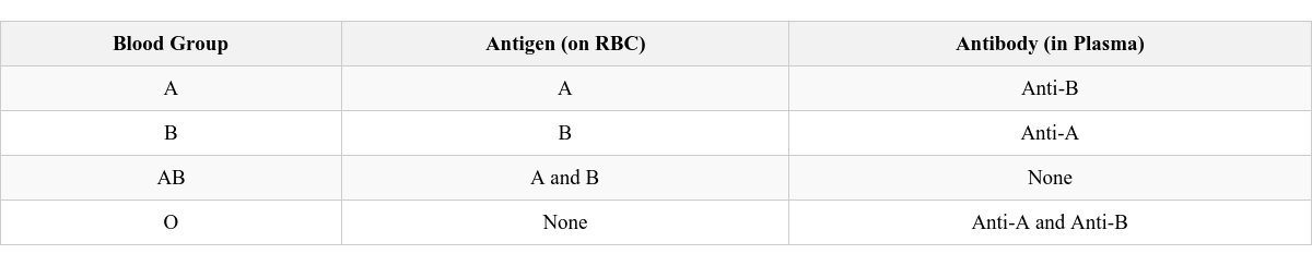
In 1900, Karl Landsteiner discovered that human beings have special types of proteins called antigens on the surface of RBCs, and other special proteins called antibodies in the blood plasma.
Two types of antigens: Antigen A and Antigen B, found on human RBCs. Based on the presence or absence of these antigens, human blood is classified into four ABO blood groups: A, B, AB, and O. Blood group A: Persons with Antigen A. Blood group B: Persons with Antigen B. Blood group AB: Persons with both Antigens A and B. Blood group O: Persons with no antigens.
Fig. 6.9 The ABO system of blood groups Antigen and Antibody Interaction
Two types of antibodies: Anti-A antibody and Anti-B antibody, found in human blood plasma. Persons with one antigen have antibodies against the other antigen. For example, persons with Antigen A (Blood group A) have Anti-B antibody (which is against Antigen B).
Clumping Reaction
Antigen A and Anti-A antibody are incompatible. Antigen B and Anti-B antibody are incompatible. When incompatible blood groups (e.g., blood from group A and group B) are mixed, clumping (or lump formation) occurs due to antigen-antibody interaction. Blood Transfusion
Blood transfusion is the transfer of blood from one person (donor) to another (recipient) in cases of severe injury or operation where there is considerable blood loss.
 Self Study
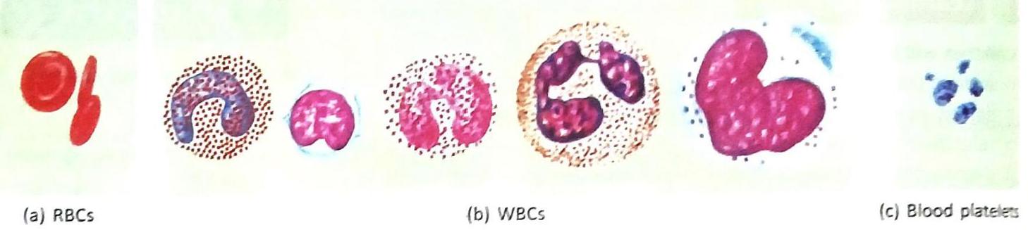

Fig. 6.2 Blood vessels
Fig. 6.2 Blood vessels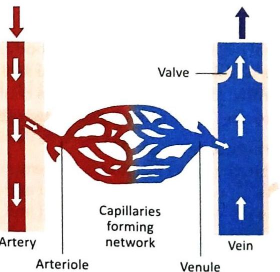
Fig. 6.4 Capillaries connect arteries and veins
Fig. 6.4 Capillaries connect arteries and veins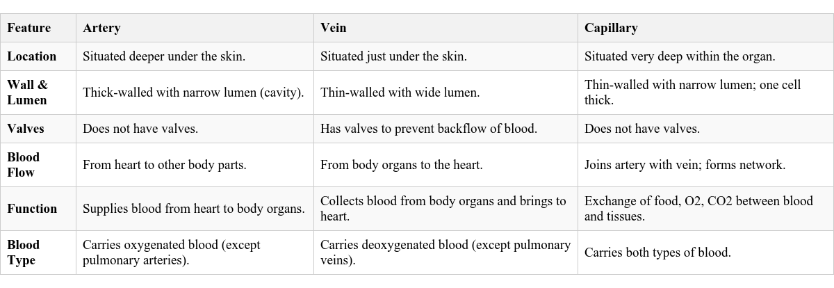

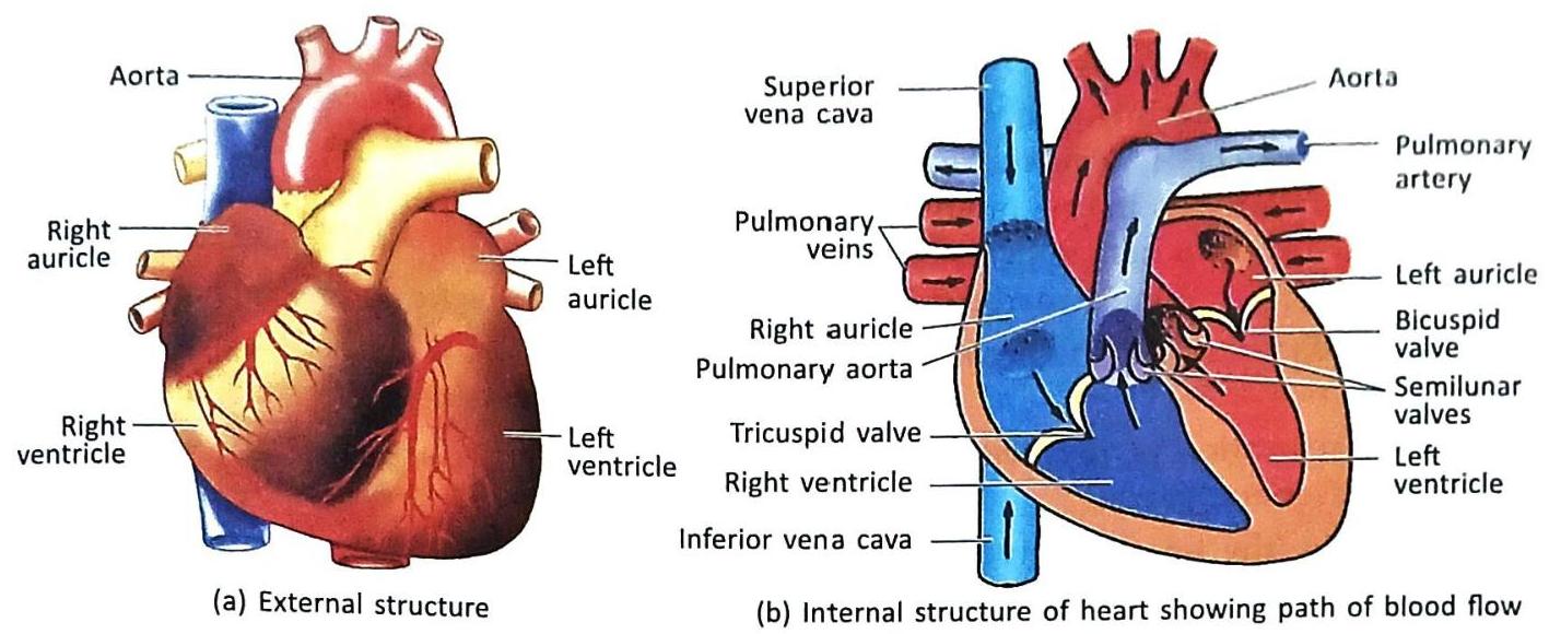
Fig. 6.5 The human heart
Fig. 6.5 The human heart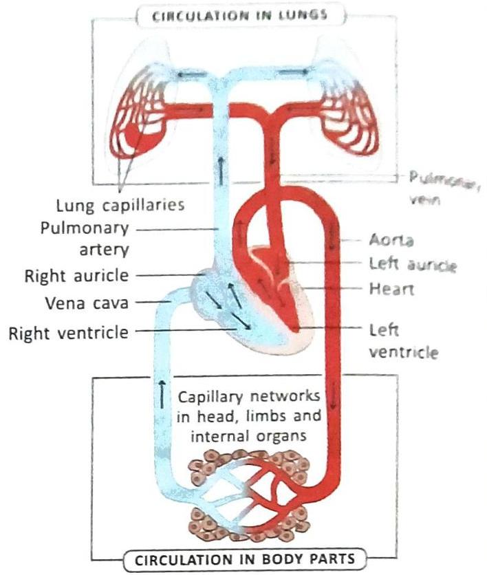
Fig. 6.6 Circulation of blood in humans
Fig. 6.6 Circulation of blood in humans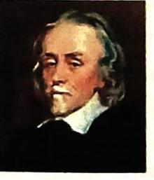
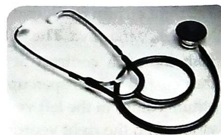
Fig. 6.8 Stethoscope
Fig. 6.8 Stethoscope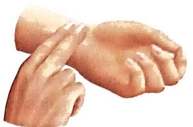

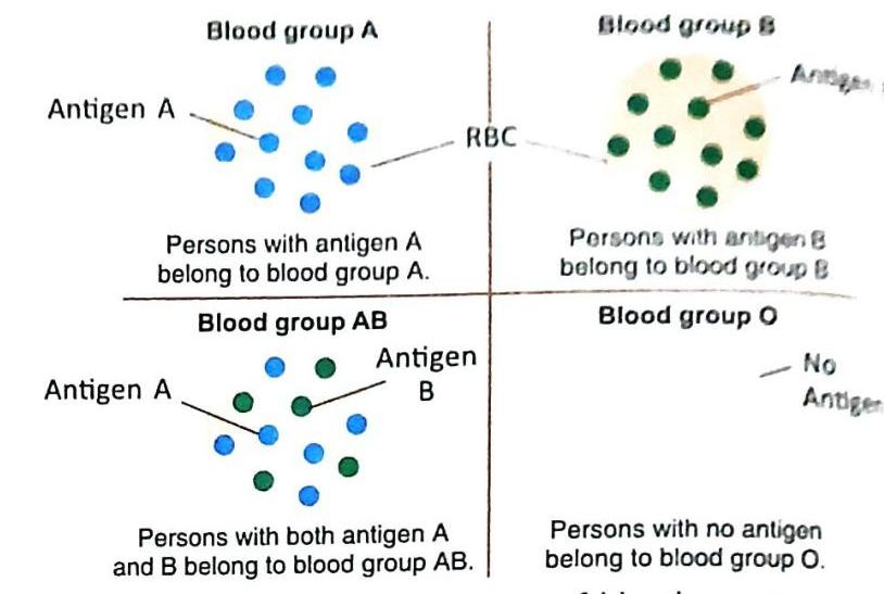
Fig. 6.9 The ABO system of blood groups
Fig. 6.9 The ABO system of blood groups