Chapter: 05. Human Respiratory System
Human Respiratory System 5
RESPIRATION
Respiration is the process by which living organisms obtain energy from food. It involves the reaction of food with oxygen to release energy.
To obtain oxygen for the oxidation of food. To release energy to carry out various life processes. Taking in oxygen by the cell. Oxidation (breakdown) of food (glucose) using oxygen. Release of energy and carbon dioxide. Elimination of carbon dioxide. It is a physicochemical process.
HUMAN RESPIRATORY SYSTEM
The human respiratory system is a complex network of organs and passages that facilitates the exchange of gases between the body and the environment. Lungs are the primary organs for respiration.
Main Respiratory Organs: Lungs Connection to External Air (Pathway): Nose
Entry Point: Air is breathed in through the nose or nostrils. Nasal Passages: Nostrils open into these passages. Functions of Nasal Passages: Air is filtered: Hair in passages traps dust, smoke, pollen, and microbes. Air is warmed: Warmed up to body temperature. Air is moistened: Becomes moist. Benefit: Ensures clean, filtered, warmed, and moistened air enters the lungs. Cause: Unwanted particles not trapped by hair irritate the sensory lining of nasal passages. Result: Unwanted particles are forcefully thrown out. Pharynx
Passage Type: Common passage for both food and air. Glottis: Opening of the trachea in the pharynx. Structure: A muscular flap. Function: Guards the glottis, closing it while swallowing food to prevent food entry into the trachea. Larynx
Location: Upper part of the trachea. Other Names: Voice box or Adam’s apple. Connection: Connects the pharynx to the trachea. Sound Production: Contains vocal cords; sound is produced by vibrations in these cords. Trachea or Windpipe
Function: Air from nasal passages reaches the lungs through the trachea. Structure: Wall supported by C-shaped rings of cartilage, which keep it open. Bronchi and Bronchioles
Bronchi (singular: Bronchus): Formation: Trachea divides into two bronchi. Entry: Each bronchus enters a respective lung. Formation: Inside the lungs, bronchi divide and redivide into smaller tubes called bronchioles. Ending: Bronchioles further divide and finally end in air sacs or alveoli. Lungs
Main Organs: Primary respiratory organs. Structure: A pair of spongy, highly elastic, and bag-like structures. Location: Enclosed in an airtight thoracic cavity. Thoracic Cavity Formation: Formed by the backbone, ribs, and sternum (breastbone). Structure: A large muscular structure. Location: Forms the floor of the thoracic cavity. Function: Separates the thoracic cavity from the abdominal cavity. Alveoli (singular: Alveolus): Structure: Numerous tiny, thin-walled, and air-filled sacs within the lungs. Number: Approximately 750 million alveoli in lungs. Surface Area: Provide a large surface area for the exchange of gases. Blood Supply: Surface is covered with a fine network of blood capillaries. During inhalation: Alveoli fill with fresh air. Oxygen: From the air in alveoli enters the blood capillaries. Carbon Dioxide: From the blood comes out into the air within the alveoli, ready to be exhaled.
Fig. 5.2 Lung alveoli associated with a bronchiole having a network of blood capillaries on their surface
MECHANISM OF RESPIRATION
Respiration is a complex process involving several steps to ensure the body receives oxygen and expels carbon dioxide while generating energy.
Differences between Breathing and Respiration
Breathing
Breathing is the physical process of moving air into and out of the lungs.
Definition: The process of taking in fresh air (rich in oxygen) and giving out used air (rich in carbon dioxide). Nature: It is a physical or mechanical process. Inhalation: Taking in air rich in oxygen into the lungs. Exhalation: Giving out air rich in carbon dioxide from the lungs. Muscles Involved: Rib muscles and diaphragm work together. Mechanism of Breathing
Ribs: Raised upwards and outwards by the contraction of rib muscles. Diaphragm: Pulled down (flattens) by contraction. Thoracic Cavity/Lungs Volume: Increases. Air Pressure (inside lungs): Decreases. Air Flow: Fresh air from the atmosphere (higher pressure) rushes into the lungs through nostrils and air passages, filling the lungs.
Fig. 5.3 Changes in chest cavity during inhalation Ribs: Move downwards and inwards (return to original position). Diaphragm: Moves upwards (returns to original position) as muscles relax. Thoracic Cavity/Lungs Volume: Decreases. Air Pressure (inside lungs): Increases. Air Flow: Air from the lungs is pushed out through air passages and nostrils.
Fig. 5.4 Changes in chest cavity during exhalation
What We Inhale and Exhale
The composition of air we inhale and exhale is different due to the process of gas exchange in the lungs.
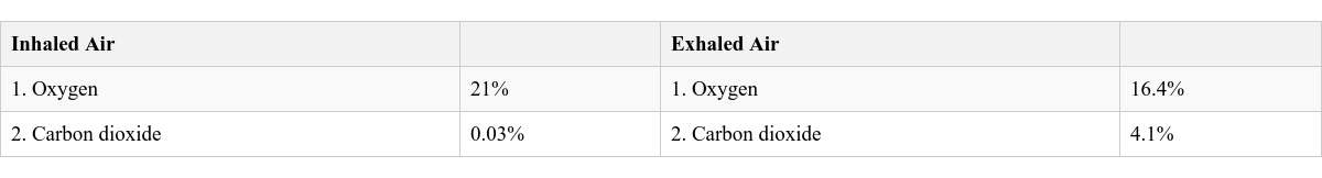
Differences between Inhaled Air and Exhaled Air
The exhaled air contains more carbon dioxide, which can be demonstrated as it turns limewater milky.
Effect of Increased Physical Activity on Breathing Rate
Breathing Rate: The number of times a person breathes (one inhalation + one exhalation) in a minute. Normal Breathing Rate: 16-18 times per minute. Effect of Physical Activity: Increases breathing rate (e.g., up to 25 times per minute during fast running and heavy exercise). Reason: Increased physical activity requires more energy, which means more oxygen is needed for oxidation of food and more carbon dioxide is produced as a waste product. The body increases breathing to meet these demands. Effect of Rest: Breathing rate slows down. Sleepiness/Drowsiness: Occurs when breathing rate slows down to an extent that insufficient oxygen is received.
External Respiration
Process: Exchange of gases between the air in the alveoli of the lungs and the blood. Enters the blood from the alveoli. Combines with haemoglobin (respiratory pigment in red blood cells - RBCs). Forms a temporary compound called oxyhaemoglobin. Oxyhaemoglobin is then carried by the blood to the body cells. From the blood, it enters the alveoli in the lungs to be exhaled. Internal Respiration
Process: Exchange of gases between the blood and the body cells. On reaching the cells, oxyhaemoglobin breaks down to release oxygen. The released oxygen from the blood enters the cells. From the cells, it enters the blood to be transported back to the lungs. Cellular Respiration or Cell Respiration
Process: The breakdown of glucose inside the cells to produce carbon dioxide and energy. This is where the actual energy release happens for cellular activities.
Fig. 5.5 Summary of respiratory process in humans
COMMON RESPIRATORY DISEASES
Asthma
Effect: Airways get inflamed (swollen and narrow) and produce extra mucus. Triggers: Coughing, wheezing, shortness of breath. Shortness of breath and difficulty in breathing. Whistling or wheezing sound while exhaling. Causes/Triggers: Allergy, occupational factors, exercise-induced, cold air, air pollutants/irritants, pollen, dust mites, mould spores, pet hair. Treatment: Cannot be cured, but symptoms can be controlled by avoiding triggers. Bronchitis
Nature: Inflammation or swelling of the bronchial tubes (which carry air to and from the lungs). Causes: Viral infection, dust, air pollution, some bacteria, smoking, occupational hazards. Pneumonia
Nature: A bacterial disease causing inflammation of the lungs, specifically affecting the alveoli. Susceptibility: Old people, infants, and young children are more susceptible. Cough with sputum (may be bloody, rusty, or green). Fast breathing and feeling short of breath. Shaking chills and fever. Chest pain during breathing and coughing. Feeling of tiredness and weakness. Diagnosis: Symptoms, blood test, chest X-ray. Treatment: Normally antibiotics, cough suppressants, and fever-reducing medicines. Tuberculosis (TB)
Nature: An infectious bacterial disease. Cause: Caused by Mycobacterium tuberculosis (MTB). Primary Target: Primarily affects lungs, but pathogens can also enter bones, brain, kidneys, etc. Coughing (sometimes with mucus or blood). Loss of appetite and weight. Diagnosis: Skin test, blood test, chest X-rays, sputum tests. Treatment: Can be completely cured with antibiotic treatment, rest, and a healthy diet. Prevention: BCG injections for children (vaccination against TB) help prevent spread.
Wrapping it Up
Respiration: A biochemical process involving gas exchange (oxygen and carbon dioxide) and the breakdown of food inside cells to release energy. Breathing: A mechanical process of pumping air in and out of the lungs. Lungs: The main human respiratory organs, located within the thoracic cavity. Breathing Mechanism: Diaphragm and ribs work together to facilitate breathing. External Respiration: Exchange of oxygen and carbon dioxide between air in the alveoli and blood. Internal Respiration: Exchange of gases between blood and body cells. Cellular Respiration: The breakdown of glucose in body cells to release energy.
KNOW THESE TERMS
 Self Study
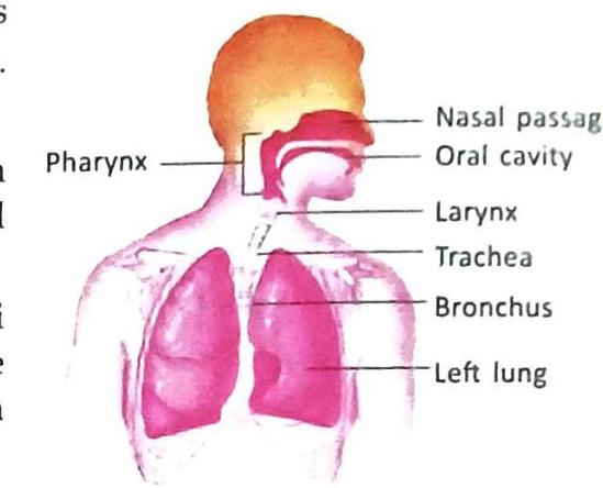
Fig. 5.1 Human lungs
Fig. 5.1 Human lungs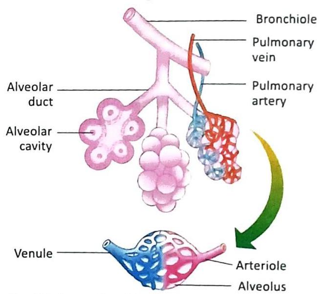
Fig. 5.2 Lung alveoli associated with a bronchiole having a network of blood capillaries on their surface
Fig. 5.2 Lung alveoli associated with a bronchiole having a network of blood capillaries on their surface

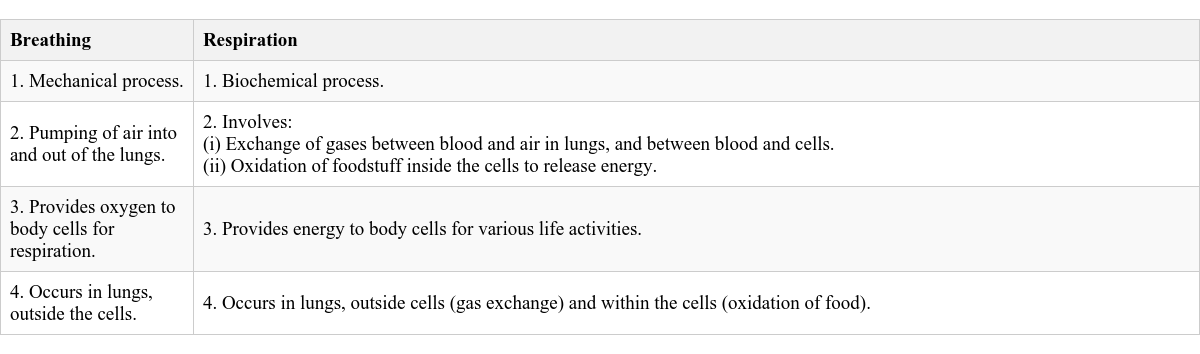

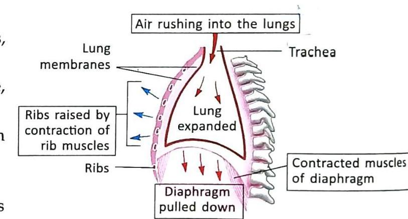
Fig. 5.3 Changes in chest cavity during inhalation
Fig. 5.3 Changes in chest cavity during inhalation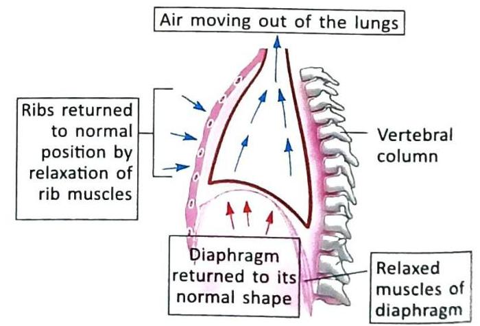
Fig. 5.4 Changes in chest cavity during exhalation
Fig. 5.4 Changes in chest cavity during exhalation

The exhaled air contains more carbon dioxide, which can be demonstrated as it turns limewater milky.
The exhaled air contains more carbon dioxide, which can be demonstrated as it turns limewater milky.

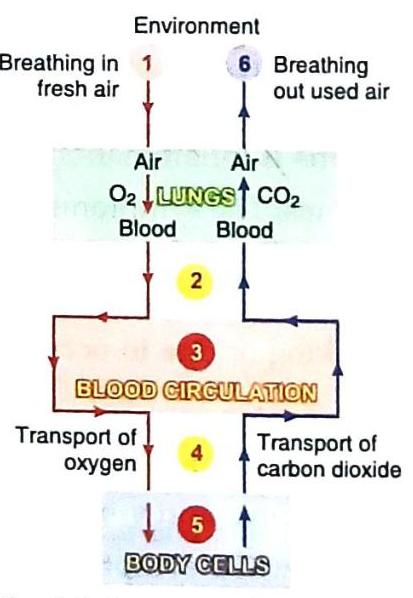
Fig. 5.5 Summary of respiratory process in humans
Fig. 5.5 Summary of respiratory process in humans