Chapter: 03. The Cell
The Cell: A Self-Study Guide for 6th Graders
WHAT IS A CELL?
Cells are the fundamental building blocks of all living things, much like bricks are the building blocks of a house. They are the smallest units within an organism that are capable of performing all the essential life functions, such as growth, metabolism (how the body uses energy), and reproduction. Therefore, cells are known as the basic unit of structure and function in all living beings.
Discovery of Cell
The discovery of cells dates back to 1665 when an English scientist named Robert Hooke observed a very thin slice of cork under a microscope he built. He noticed that the cork was made up of numerous small, empty compartments, which reminded him of the small rooms (cells) in a monastery. He named these tiny boxes ‘cells’. These ‘cells’ were actually the empty spaces left behind by dead plant cells, surrounded by their strong cellulose walls.
The detailed structure of cells and the functions of their various parts were only understood much later, with the invention of more powerful microscopes like the electron microscope. The scientific study of cells is called cytology.
Cell Theory
The concept that all living organisms are made up of cells was explained as the Cell Theory by two scientists, Schleiden and Schwann. This fundamental theory of biology states two main points:
Cells are the structural and functional units of all living organisms. New cells always arise from the division of pre-existing cells. Know Your Scientist: Robert Hooke
Robert Hooke (1635-1703) was a brilliant English scholar known as a “polymath” because he excelled in many different fields, including physics, astronomy, geology, meteorology, and architecture. He is particularly recognized for:
Hooke’s law of elasticity in Physics, which describes how springs stretch. The construction of a compound microscope, a significant advance in scientific instrumentation. His famous observation of cork under the microscope, leading to the discovery of cells. Coining the term ‘Cell’ to describe the basic unit of life. Because of his wide-ranging contributions, he is often described as a “jack of all trades.”
CELLS - THEIR NUMBER, SHAPE AND SIZE
Cells show incredible variety in their number, shape, and size, which are often related to the specific functions they perform.
Number of Cells
Based on the number of cells they are made of, organisms are classified into two main types:
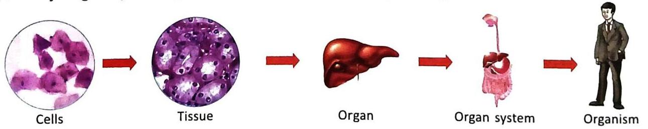
Unicellular Organisms (uni = single + cellular = celled): These organisms are formed of just one single cell. Their single cell performs all the life processes necessary for survival. Examples of unicellular plants include Bacteria, Chlamydomonas , and Yeast. Examples of unicellular animals include Amoeba and Paramecium. Multicellular Organisms (multi = many + cellular = celled): These organisms are formed of many cells, ranging from millions to billions. Most plants and animals we commonly see around us are multicellular, such as Peepal trees, Neem trees, Rose plants, Earthworms, Fish, Frogs, and Humans. In multicellular organisms, different cells become specialized to perform specific functions more efficiently. Cells that are similar in structure and dedicated to performing a specific function group together to form a tissue. Several tissues work together to form an organ (e.g., heart, liver). Different organs, each specialized for a particular function, join together to form an organ system (e.g., digestive system, circulatory system). Finally, many organ systems work together to form a complete organism (e.g., a human being). Shape of Cells
Cells exhibit a wide variety of shapes, which are directly related to the specific functions they perform. Cells can be:
Let’s look at some examples:
Guard cells of stomata (tiny pores on leaves) are kidney-shaped or bean-shaped cells. Mesophyll cells of leaves are long and rectangular, packed with chloroplasts for photosynthesis. Xylem vessels in the vascular bundles (transport tissues) of roots and stems are cylindrical cells, forming tubes for water transport. White blood cells (WBCs) are amoeboid, meaning they can change their shape and move like an Amoeba by extending temporary projections called pseudopodia (pseudo = false, podia = feet). Muscle cells are spindle-shaped (tapering at both ends) and help in movement. Nerve cells (neurons) are quite long and have branched ends, allowing them to transmit messages over long distances. Fat cells, liver cells, red blood cells (RBCs) , and egg cells are typically rounded or spherical. Size of Cells
Cells also vary greatly in size.
The smallest cells are often bacteria, which can be as tiny as 0.1 to 0.15 microns (a micron is one-thousandth of a millimeter). The longest cells can be as long as one meter, such as certain nerve cells. In the human body, red blood cells (RBCs) are among the smallest at about 7.0 microns, while nerve cells are the longest, potentially over a meter long. The unicellular alga Acetabularia is unusually large for a single cell, measuring about 10 cm long. The egg of an Ostrich is the largest known single cell, with a diameter of about 15-20 cm. It’s important to remember that larger organisms do not necessarily have bigger cells; instead, they have a greater number of cells.
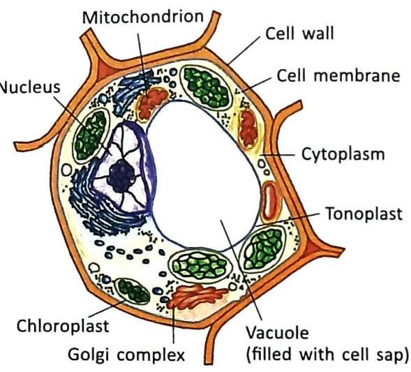
CELL STRUCTURE
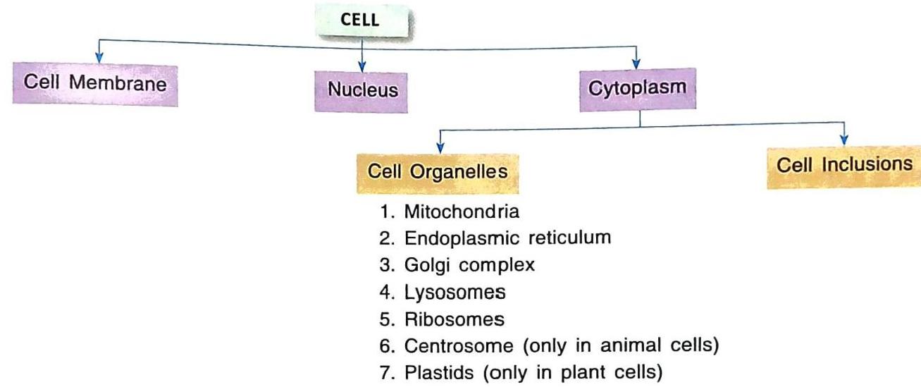
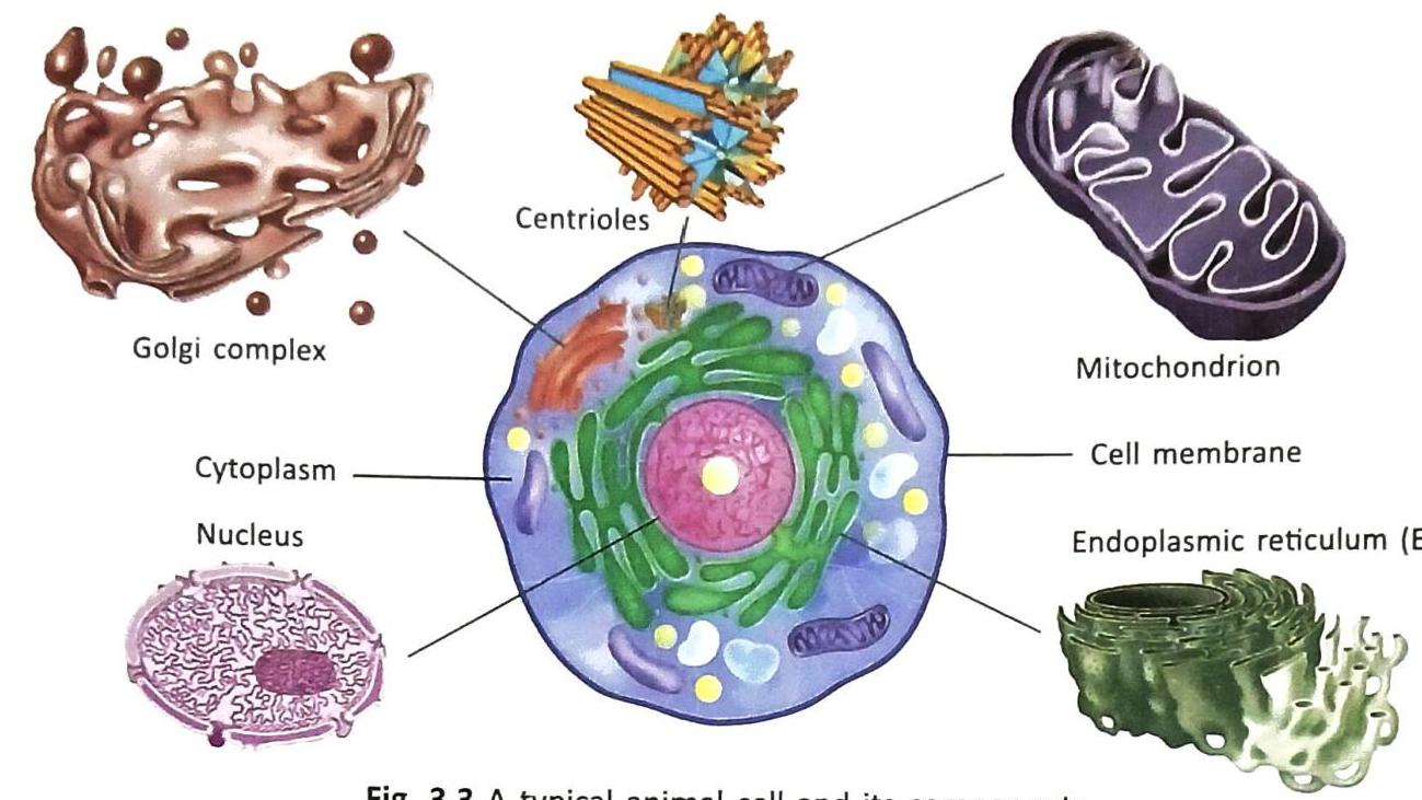
A typical cell is composed of three fundamental parts: the Cell membrane, Cytoplasm, and Nucleus. These components work together to ensure the cell carries out its life processes.
Cell Membrane
The cell membrane, also known as the plasma membrane, is a thin, flexible outer covering that surrounds the cytoplasm of the cell.
Functions of the Cell Membrane:
It forms the outer boundary of the cell. It separates one cell from another, and also separates the cell’s internal environment from its external surroundings. It helps to maintain the specific shape of the cell. It acts as a gatekeeper, allowing only certain substances (like oxygen, water, and nutrients) to enter and leave the cell, while preventing others. For this reason, it is called a selectively permeable membrane. Cell Wall
The cell wall is an additional outer layer found only in plant cells, located outside the cell membrane. It is a non-living structure primarily composed of cellulose. Unlike the cell membrane, the cell wall is permeable, meaning it allows both liquid and gaseous substances to pass through freely.
Functions of the Cell Wall:
It provides rigidity and a definite shape to the plant cell. It offers mechanical support, helping the plant stand upright and resist pressure. It protects the internal organelles of the cell from external damage. Nucleus (Control Centre of Cell)
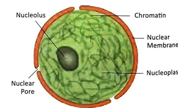
The nucleus is a prominent, usually spherical or oval structure within the cell. It was discovered by Robert Brown in 1831. The nucleus is like the brain of the cell, controlling all its activities.
The nucleus consists of four main parts:
Nuclear envelope: This is a double-layered membrane that surrounds the nucleus. It has many small openings called nuclear pores, which allow substances to move in and out of the nucleus. Nucleoplasm: A thick, fluid-like substance that fills the nucleus. Nucleolus: A small, rounded body located inside the nucleoplasm. It plays a crucial role in forming ribonucleic acid (RNA), which is essential for the production of ribosomes. Chromatin material: This appears as thread-like structures made of nucleo-protein fibres (called chromatin fibres). In a non-dividing cell, they form a network within the nucleoplasm. Something More About the Nucleus:
Chromatin fibres are made up of deoxyribonucleic acid (DNA) and proteins. DNA is the hereditary material, meaning it carries the genetic instructions that make each organism unique and are passed from parents to offspring. During cell division, these chromatin fibres condense and become tightly packed to form thick, darkly stained structures called chromosomes. Every cell in the human body normally contains 46 chromosomes (which is 23 pairs). Chromosomes are typically visible only when a cell is actively dividing. The number of chromosomes varies significantly from one species to another. Important Notes on Nucleus Presence:
Normally, most cells have only one nucleus, making them uninucleate. However, some cells, like striated muscle cells, can contain more than one nucleus, making them multinucleate. Interestingly, mature red blood cells (RBCs) in the human body are unique because they do not have a nucleus; they are enucleate. Functions of the Nucleus:
The nucleus controls all the activities of the cell, earning it the nickname “control centre of the cell.” It stores and transmits genetic information for hereditary characteristics from parents to their children. This is why children often resemble their parents. It plays a vital role in cell division, ensuring new cells are formed correctly.
Cytoplasm
The cytoplasm is a jelly-like, semi-fluid substance that fills the cell, located between the cell membrane and the nucleus. It is primarily composed of water, up to 90%. Many vital life processes and chemical reactions of the cell take place within the cytoplasm. It is also where a number of specialized structures, called cell organelles, are suspended.
PROTOPLASM
Protoplasm is the collective term for all the living matter within a cell. It includes both the nucleus and the cytoplasm, all enclosed by the cell membrane. It is considered the “living substance” of the cell.
Cell Organelles
Suspended within the cytoplasm are various living structures known as cell organelles. Each organelle has a specific structure and performs a particular function, contributing to the overall life of the cell. Some key cell organelles include:
Plastids (found only in plant cells) Centrioles (found only in animal cells) Plastids
Plastids are a special type of organelle found only in plant cells. They come in three main types, each with a different role:
These are green-coloured plastids because they contain a green pigment called chlorophyll. Chlorophyll is essential for photosynthesis, the process by which plants use sunlight to make their own food. Chloroplasts give the green color to plant leaves and are often called the “kitchen of the cell” because food is prepared there. These are coloured plastids that contain pigments of red, yellow, and orange colours. They are responsible for giving the varied vibrant colours to flowers and fruits. These are colourless plastids. Their primary function is to store starch, proteins, and fats as reserve food materials. Vacuoles
Vacuoles are clear, fluid-filled spaces found within the cytoplasm. Their appearance and function differ between plant and animal cells:
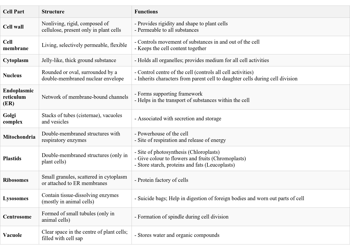
Mature plant cells typically have a single, large central vacuole that can take up a significant portion of the cell volume. Young plant cells may have many small vacuoles that eventually merge to form a large one. The cavity of the vacuole is filled with a liquid called cell sap. The vacuole is separated from the cytoplasm by a membrane called the tonoplast. Functions in plant cells: Vacuoles help keep the plant cell turgid (stiff and firm) by maintaining internal pressure, which provides support to the plant. The cell sap within the vacuole stores various substances like food, water, pigments, and waste products. Animal cells are either without any vacuole or have a few small, temporary vacuoles (such as food vacuoles or contractile vacuoles in single-celled organisms like protozoans). These are not permanent large structures like in plants. It’s important to remember that only plant cells have a cell wall. Table 3.1 Cell Parts, their Structures and Functions
This table summarizes the key components of a cell, their structural characteristics, and their primary roles:
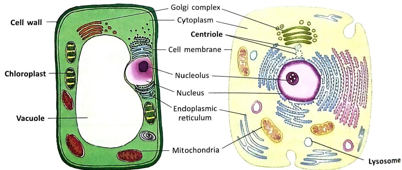
PLANT CELLS VERSUS ANIMAL CELLS
While both plant and animal cells are eukaryotic (have a nucleus and membrane-bound organelles), there are some distinct structural differences. Certain structures are found exclusively in one type of cell and not the other.
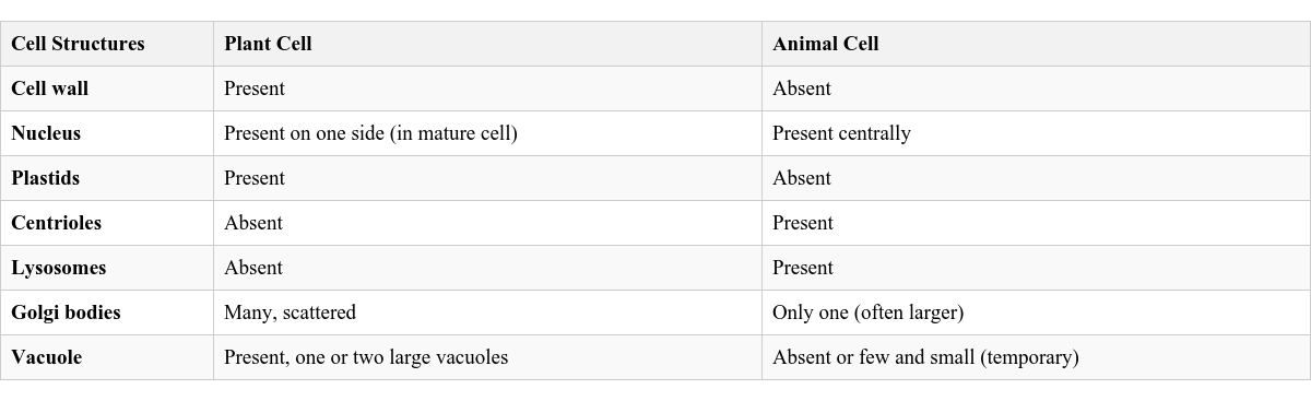
Table 3.2 Differences between Plant and Animal Cells
Need of Cell Division
Cell division is a fundamental process in all living organisms where new cells are formed from existing ones. This process is essential for several critical reasons in our body:
Growth: New cells are continuously produced, which allows our bodies to grow from a small child into an adult. Repair Wounded Tissues: When we get a cut or injury, cell division helps to replace the damaged cells and repair the wounded area. Replace Old and Worn Out Cells: Cells in our body have a limited lifespan. Cell division ensures that old or damaged cells are constantly replaced by new, healthy ones, keeping our tissues and organs functioning properly. Thus, cell division is a necessary and continuous event in the life of all living beings, ensuring their survival, growth, and repair.
HOW TO STUDY CELLS
Cells are generally too small to be seen with the naked eye. Therefore, they are studied using a microscope, which magnifies their image. To make various parts of the cell more visible and identifiable, scientists often color the cells with special dyes. This process of coloring is called staining, and the dye used is called a stain. Once stained, the cells are typically placed on a clean glass slide and observed under the microscope.
Here’s what you might observe when studying different types of cells:
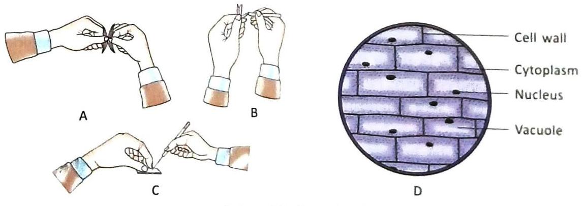
Studying Plant Cells (Onion Peel)
When a thin layer of onion peel is stained and observed under a microscope, you can see:
Brick-shaped cells arranged neatly side-by-side. Each cell has a distinct, darkly stained cell wall as its outer boundary. A large, centrally placed vacuole is usually visible, taking up a significant portion of the cell. A thin layer of cytoplasm is seen between the cell wall and the vacuole. The nucleus might be visible, often pushed to one side by the large vacuole.
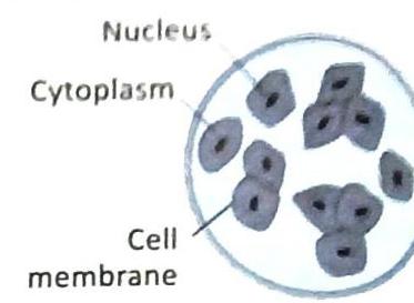
Studying Animal Cells (Human Cheek)
If you gently scrape the inside of your cheek and observe the cells under a microscope after staining, you would notice:
A large number of scattered cells, not arranged in a rigid pattern like plant cells. Each cheek cell is typically polygonal or irregular in shape, lacking a fixed, rigid outline. A darkly stained nucleus is usually visible near the center of the cell. A thin plasma membrane (cell membrane) surrounds the cell cytoplasm. Vacuoles and a cell wall are absent in these animal cells.
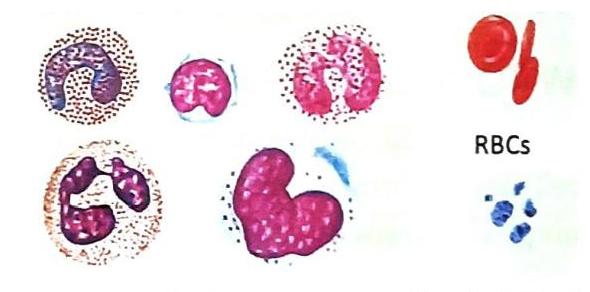
Studying Blood Cells
Observing a blood sample under a microscope reveals different types of blood cells:
White blood cells (WBCs): These appear as amoeboid (irregularly shaped) cells with a distinct, often lobed nucleus. Red blood cells (RBCs): These are concave (like a disc with a depressed center) and do not have a nucleus. They are typically numerous and biconcave discs. Blood platelets: These are very small and often difficult to see clearly under a standard compound microscope.
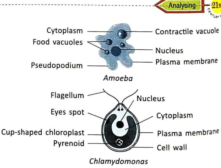
Studying Unicellular Organisms (Amoeba and Chlamydomonas)
When observing slides of single-celled organisms like Amoeba and Chlamydomonas:
Its body is irregular in shape and constantly changes by extending temporary, finger-like projections called pseudopodia, which it uses for movement and feeding. The cytoplasm is jelly-like, dense, colourless, and transparent. It contains a single contractile vacuole (for water regulation) and many small food vacuoles (for digestion). A single circular nucleus is present in the center of the cytoplasm. Its body is simple, unicellular, and typically spherical or ovoid. The cytoplasm is surrounded by a plasma membrane and an outer cell wall (like a plant cell). It has a prominent cup-shaped chloroplast containing a single pyrenoid (the site where starch is formed). Two whip-like structures called flagella are present at one end, used for locomotion. A light-sensitive organ called a stigma or eyespot helps it detect light. The nucleus is typically enclosed by the large chloroplast.
KNOW THESE TERMS
Cell: The basic unit of structure and function of all living organisms. Microscope: An instrument that makes the enlarged image of an object. Electron microscope: A microscope that makes a highly enlarged image by the interaction of electrons with the object. Micron: A unit of length equal to one thousandth part of a millimeter, i.e., 0.001 mm.  Self Study